
3. The Blastula
Free content. Always.
3. The Blastula
Of course, that is not actually a photo of Jupiter on the cover. It is a picture of a blastula, curtesy of Sindelka and colleagues (2017).
We briefly discussed the blastula in lecture 1. As a reminder, the blastula is formed after the initial divisions of embryonic cells. It is essentially a "blob" of many embryonic stem cells. Again, here is the embryonic life cycle for reference:

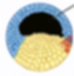
And here is just the blastula:

To clarify--In the cover photo (the "Jupiter" blastula), the color of the blastula is graded (darker on top, lighter on bottom). This is because cells tend to be denser at the top (the animal pole), and scarcer at the bottom (the vegetal pole) due to the formation (or preparation for formation) of the yolk at the animal pole. This gradient becomes more clear as the blastula continues to develop. A slightly more accurate cartoon representation of a blastula would actually be this one:

The first illustration isn't necessarily incorrect; the second illustration is just depicting a blastula a bit further along in development, when the poles are very distinguishable. A good way to remember the poles is this: the animal pole forms the yolk sac (hence the animal), the vegetal pole provides nutrition to the animal (think vegetal -> veggies -> nutrition). Animal pole on top, vegetal pole on bottom.
If we were to slice the blastula in half, we would find a space in the center, near the animal pole. This space is called the blastocoel (pronounced BLAST-OH-SEAL). Here's a close-up, again extracted from the embryonic life cycle illustration:

The blastocoel is a crucial component of early development, because it creates necessary space for cell movement and embryonic changes. And as we will learn, cells move in a conserved, guided manner to form the developed body and nervous system. As little preview: cells move from the outside of the blastula into the blastocoel and coats its inner perimeter through a process called gastrulation. But do not worry about this process yet, focus on just the blastula for now.
We've discussed cell fate in the previous lecture. At the blastula stage, all cells are stem cells and can develop into any part of the body later on. Modern biotechnology has allowed us to track cell development of the blastula and determine the ultimate cell fate of blastula cells. Here's how fate mapping works:
inject different cells with differently colored dye
allow the blastula to continue to develop
track where stained cells end up in the end

So, even though cells in the blastula are considered stem cells with vast cell potential, systematic biochemical signaling throughout development gradually determines more and more of a cell's fate. Weird to think about. It's about to get a whole lot weirder. - Feel free to take a break here -
After a little bit more development, the blastula enters a stage called the mid-blastula transition. Importantly, in this stage, the cells in the embryo begins using its own genome for RNA transcription (i.e., the making of proteins), therefore regulating its own gene expression. Because of this pivotal change to self-governed gene regulation, previously totipotent blastula cells are becoming pluri- and multi- potent, such that they become the broad categories of ectoderm cells (blue), mesoderm cells (red), and endoderm cells (yellow):
Of course, these cells are not actually color-coded in real life and all look pretty much identical to the naked eye. They mostly still look like this:

So how do we know that cells begin to differentiate into the ectoderm, mesoderm, and endoderm at this stage? Neuroscientists, particularly Spemann and Mangold, came up with an incredibly clever way of distinguishing the three cell layers at this stage, which we will dedicate the next lecture to.
As always, I hope you learned something new (even if it is a factoid) and are finding developmental neurobiology interesting (even if just a little bit more). Hope to see you back.
Neurocookie.org believes that neuroscience should be fun, accessible, interesting, jargon-free, and welcoming to all who wishes to learn, no matter your background.
Your Instructor
Cocoa (Cookie)

Cookie the science cat.