
3.5. Spemann and Mangold's Organizer
Free content. Always.
3.5. Spemann's Organizer
In our previous lecture, The Blastula, we discussed how totipotent blastula cells gradually reduce in potency, such that they are now categorized into three cell types: ectoderm cells (blue), mesoderm cells (red), and endoderm cells (yellow):
The general layout of the three cell groups are determined at this stage, with ectoderm cells at the animal pole, endoderm cells at the vegetal pole, and mesoderm cells at the dorsal pole, in-between ectoderm and endoderm cells. According to our fate map of the blastula, here's what each of the three cell types generally become:

Remember that cell fate maps are made retroactively through the fate mapping technique. Notice that the ectoderm cells eventually become both the epidermis and nervous system in the fully developed body. Specifically, ectoderm cells on the ventral side develop into the epidermis, and ectoderm cells on the dorsal side develop into the nervous system. So, how does the ectoderm know whether to become the epidermis or the nervous system? Turns out, close contact of the ectoderm with the mesoderm (at the dorsal pole) turns ectoderm cells into the nervous system. But what's the mechanism?
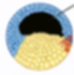
Let's first zoom in to the late blastula (after the mid-blastula transition stage), where we will find a small "hump" between the mesoderm (red) and the endoderm (yellow) that is not shown in the graphics above. This small "hump" is called the blastopore lip, visibly shown below:

Do not worry about labels shown in the picture; just know that the little "hump" between the mesoderm and endoderm is the blastopore lip. As a preview, in the next stage of embryonic development, the gastrula stage, cells on the surface of the embryo begin to migrate inward, through the blastopore lip. But let's remain on the blastula for now.
The blastopore lip is mostly composed of mesoderm cells. Today, know that the mesoderm induces the ectoderm to become neural tissue. But long ago, scientists only had a hunch about this--they weren't completely sure. Their twisted, curious minds wondered: if we take out the blastopore lip (from the dorsal side of cell a.) and transplant it onto the ventral side of a completely different cell at the same developmental stage (cell b.), what will happen?

Specifically, what will happen to cell b., which now has two blastopore lips--one it developed naturally on the dorsal side, and one that was transplanted from cell a. onto its ventral side? Neuroscientists (and maniacs) Spemann and Mangold conducted exactly this experiment on frog embryos. They found that if cell b. continues its development, the embryo will develop two neural axes--it will literally grow two heads.

Discovery of this freaky little creature won Spemann and Mangold the Nobel Prize in 1935. More importantly, it confirmed that the mesoderm signals the ectoderm to become neural tissue via proximity. So what are the signals? You'll have to wait to find out.
As always, I hope you learned something new (even if it is a factoid) and are finding developmental neurobiology interesting (even if just a little bit more). Hope to see you back.
Neurocookie.org believes that neuroscience should be fun, accessible, interesting, jargon-free, and welcoming to all who wishes to learn, no matter your background.
Your Instructor
Cocoa (Cookie)

Cookie the science cat.